Coordinates in the Brain
UK, Medical Research Council (MRC), Cognition and Brain Sciences Unit (CBU), Internet
Reporting activity or lesion location
Chris Rorden and Matthew Brett
[paraphrase]
Stereotaxic coordinates
MRI scans from multiple individuals will vary greatly due to differences in slice orientation and brain features (i.e. brain size and shape varies across individuals). Therefore, it is generally useful to ' normalize ' scans to a standard template. Normalization is the process of translating, rotating, scaling, and maybe warping a brain to roughly match a standard template image. After normalization, it may be informative to report locations using stereotaxic ("Talairach") coordinates. This format uses three numbers (X,Y,Z) to describe the distance from the Anterior Commissure (the 'origin' of Talairach space). The X,Y,Z dimensions refer to left-right, posterior-anterior, and ventral-dorsal respectively. So 38x-64x58mm refers to a point in right posterior dorsal region of the brain. If you have normalized your brain to one matching the brain in the Talairach atlas, then you can examine the Talairach atlas to get a general idea of the region you are describing. Because most neuroimagers coregister their images with a template from the Montreal Neurological Institute, readers can also examine an MNI template (as there are some differences between the MNI brain and the Talairach brain ). The next section will describe some limitations in making comparisons between brains.
Limitations of Comparisons
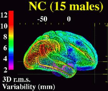
Like fingerprints, each brain has a unique configuration of gyri and sulci. In fact, secondary and tertiary sulci are not found in all individuals. This makes it difficult and probably undesirable for software to precisely match sulci between different individuals. In addition, because the sulci can have very different configurations, software which attempted to match sulci would create severe local distortions. Therefore, most normalization software smooths brain images before attempting normalization. The detail of sulcal information is greatly reduced by this smoothing process. Therefore, the normalized images for any two individuals will not have the sulci and gyri in the same locations. As a consequence, stereotaxic coordinates do not necessarily refer to a specific sulcal location. In this way, stereotaxic coordinates are 'probabilistic.’ A Figure shows the variability of sulcal location after normalization in 15 healthy individuals, after a normalization based on matching the location of the major sulci. Also, note that brain function may not be specifically located to specific sulci. So sulcal location and stereotaxic space may both be probabilistic in terms of functional location. In a study of cortical cytoarchitecture, researchers noted that "sulci are not generally valid landmarks of the microstructural organization of the cortex". Unfortunately, the cytoarchitecture cannot be directly examined in living humans. Therefore stereotaxic coordinates and sulcal landmarks are the best we can do.
Identifying Brodmann's Areas
Brodmann (1909) classified brain regions based on their cytoarchitecture (basically, the appearance of the cortex under the light microscope). In some instances there is a clear link between the microscopic appearance of a region and its function. For example, the 'stripe' of the striate cortex delineates the first main cortical area of the visual system (today this area is usually referred to as V1, Brodmann called it area 17). However, it is important to remember that Brodmann's Areas (BAs) were identified purely based on visual appearance, which is not necessarily related to function. Attempting to convert stereotaxic coordinates based on MRI scans to Brodmann areas based on cytoarchitecture faces several problems. First, as noted earlier, Zilles et al. have found that there can be only approximate correspondence between sulcal landmarks or stereotaxic space and cytoarchitecture. Second, while many atlases define BAs (somewhat arbitrarily) as being segmented at sulcal fundus, sulcal location varies tremendously between individuals (see the previous section). Atlases such as the online Talairach Daemon and Van Essen and Drury's Caret software can approximate between stereotaxic location and BAs, although the derivation of the BAs can be obscure, and in neither case is based on cytoarchitectonic data from the individual brains. These tools should be used with extreme caution, because of the inherent uncertainty of the information they contain. Given the problems with these BA estimates, it is attractive to take a more conservative approach, and define locations based on stereotaxic coordinates or (where activity across individuals appears to be correlated more strongly with sulcal landmarks than Talairach space) to sulcal locations. The main reason for converting between stereotaxic space and BAs is when comparing human work to animal research (where direct stereotaxic comparisons are impossible).
Locating Sulci and Gyri
It is important to remember that neither stereotaxic coordinates nor cytoarchitecture perfectly correspond with gyri and sulci. For reporting data from stereotaxically aligned images, it is generally a good idea to present both stereotaxic coordinates and an image, so that readers can identify relevant landmarks. Once MRI scans have been normalized, they may roughly match the Talairach atlas or the online Talairach Daemon, and you can refer to these sources to locate the relevant sulci, gyri, and (with a lower degree of accuracy) the Brodmann Area. As noted earlier, some brains do not show all of the sulci, and there are different sulcal configurations. Therefore, care should be taken when identifying a sulcus.
[end of paraphrase]


