
|
Scientific Understanding of Consciousness |
|
Sensory-Motor Cortex Speech Map
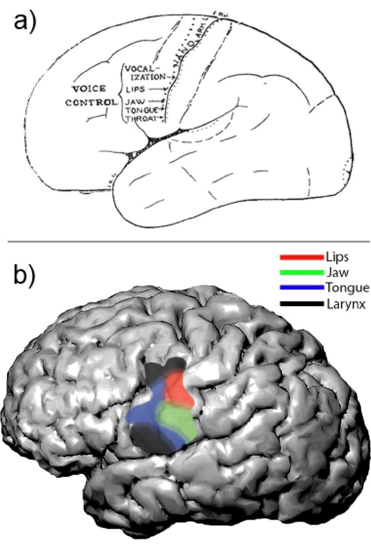
Current Opinion in Neurobiology, Volume 24, February 2014, Pages 63–67 Speech Map in the Human Ventral Sensory-Motor Cortex David Conant, et.al. Departments of Neurological Surgery and Physiology, University of California, San Francisco [paraphrase] The spatial maps of the ventral sensory-motor cortex (vSMC) are arranged in a somatotopic fashion within ventral SMC. This region has unique features and connectivity that may give insight into its specialized function in speech production. New methods allow us to probe further into the functional role by studying the spatial dynamics of vSMC during natural speaking in humans. Speaking is a unique and defining human behavior. It is carried out by precise, controlled movements of different articulators of the vocal tract, which are closely coordinated with the larynx and respiration. Speech articulation is often described as the most complex motor behavior because over 100 muscles are involved, and the movements occur on an extremely rapid time scale. Despite its complexity, nearly all of us learn to master this skill to speak fluently and effortlessly. A key brain area in the neural control of articulation is the ventral portion of the sensory-motor cortex (vSMC). Injuries to this area produce motor deficits in articulation, called dysarthria. In comparison to the dorsal sensorimotor cortical regions involved in arm reaching and hand function, the neurobiology of vSMC is relatively understudied. The vSMC features some important anatomic and functional differences from dorsal sensory-motor cortex, while sharing others. For example, in contrast to the dorsal areas, vSMC projects via the corticobulbar tract to the oro-facial motor nuclei, and ultimately to the articulatory muscles. vSMC has connections with higher-order cortical areas such as the anterior cingulate and supplementary motor area, basal ganglia, and cerebellum. With the advent of functional imaging such as PET and fMRI it became possible to noninvasively study humans during vocalization, with enough spatial resolution to investigate somatotopic maps. These studies have generally recapitulated the stimulation findings about the cortical representation of the lips, jaw and tongue. [end of paraphrase]
Return to — Auditory System Return to — Language Return to — Movement Control
|